Феномен Рейно (ФР) представляет собой эпизоды преходящей дигитальной ишемии вследствие вазоконстрикции дигитальных артерий, прекапиллярных артериол и кожных артериовенозных шунтов под влиянием холодной температуры и эмоционального стресса [1]. В 1862 г. Maurice Raynaud впервые описал этот феномен в виде кратковременного и обратимого изменения цвета кожи под воздействием холода и назвал локальной асфиксией конечности [2].
Распространенность ФР в популяции в среднем составляет 3–21 %, но данные могут варьироватьcя в зависимости от стран, что связано с разными климатическими условиями [3]. Низкая температура, стресс, беспокойство, зрелый возраст и женский пол – факторы, определяющие тяжесть и частоту вазоспастических реакций (атак Рейно).
Патогенез этого феномена изучен недостаточно. Предположительно в его основе находится дефект центральных и локальных механизмов регуляции сосудистого русла, приводящий к чрезмерно выраженному вазоспазму в ответ на раздражители. Также имеет значение изменение эндотелиальных клеток и реологических свойств крови.
Классический вариант ФР представляет собой трехфазное изменение цвета кожи: в результате вазоспазма наблюдается побеление, из-за развития гипоксии сменяющееся на цианоз. Позже вследствие реактивной гиперемии происходит покраснение кожных покровов. Таким образом, для ФР характерно трехфазное изменение кожных покровов пальцев, однако последние две фазы (цианоз и покраснение) могут возникать не всегда, поэтому нужно помнить, что для диагностики ФР достаточно посинения пальцев. При ФР в основном поражаются пальцы кистей: изменение цвета начинается с кончика пальца, распространяется на 1, 2, 3 фаланги или другие пальцы и становится симметричным на обеих руках. Зачастую ФР сопровождается онемением, покалыванием, болью, возможно изменение окраски носа, языка, ушей.
ФР подразделяют на первичный и вторичный. Первичный ФР (идиопатический) встречается в 90% случаев, возникает в более молодом возрасте по сравнению со вторичным, в 9 раз чаще встречается у женщин [4]. Обычно в этом случае наблюдается симметричное поражение 2–4 пальцев кистей. Это состояние имеет благоприятный прогноз, поскольку для него трофические изменения и прогрессирование не характерны [5].
Вторичный ФР ассоциирован с аутоиммунными заболеваниями (системная склеродермия, системная красная волчанка, дермато-, полимиозит, синдром Шегрена и др.), инфекционными заболеваниями (гепатиты В и С, микоплазменные инфекции), эндокринными (акромегалия, микседема, сахарный диабет, феохромоцитома), гематологическими (пароксизмальная ночная гемоглобинурия, полицитемия) заболеваниями, с приемом некоторых лекарств (оральные контрацептивы, алкалоиды спорыньи, бромокриптин, бета-блокаторы, цитостатики, интерферон альфа) и может быть в составе неопластического синдрома при лимфомах, лейкозах, миеломе и др. Вторичный ФР поражает чаще мужчин старше 30 лет и характеризуется асимметричными эпизодами вазоспазма с ишемическими повреждениями кожи [6].
Недавние исследования показали, что ФР впервые может возникнуть за 20 лет до развития клиники системного заболевания [7]. Исходя из этого необходимо помнить о важности ранней диагностики ФР, определения причины его возникновения, поскольку это влияет на дальнейшую тактику ведения пациентов.
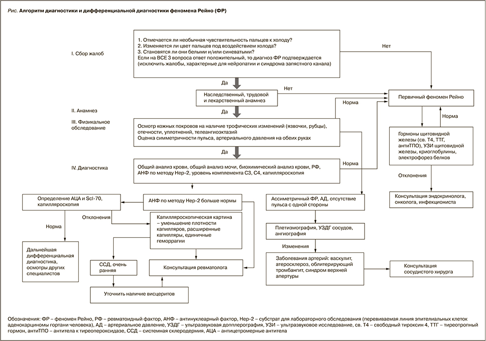
Врачам следует уделить особое внимание сбору жалоб и выявлению клинических симптомов. Диагноз ФР считается достоверным при положительном ответе на следующие вопросы [1]:
- Отмечается ли необычная чувствительность пальцев к холоду?
- Изменяется ли цвет пальцев под воздействием холода?
- Становятся ли они белыми и/или синеватыми?
Однако нужно помнить, что чувствительность пальцев к холоду встречается и у здоровых людей, поэтому подробно следует расспросить о следующем:
- Симметричность, длительность и частота возникновения поражения.
- Сопровождается ли онемением, покалыванием, болью.
- Отмечается ли побеление других участков тела (кончик носа, уши).
Второй шаг – подробный сбор анамнеза.
- Наследственный анамнез: имеются ли схожие проявления у родственников первой линии?
- Трудовой анамнез: связана ли работа пациента с воздействием вибрации или других механических факторов, травмирующих кисти?
- Лекарственный анамнез: принимает ли больной какие-либо лекарственные средства (бета-блокаторы, цитостатики, оральные контрацептивы, интерферон-альфа)?
Дальнейшее физикальное обследование направлено на выявление любых обструктивных или воспалительных заболеваний сосудов и на поиск симптомов ассоциированных аутоиммунных состояний. Осматривают кожу на наличие трофических изменений (язвочки, рубцы), отечности, уплотнений, телеангиэктазий. Особое внимание следует обратить на симметричность пульса, артериального давления на обеих руках. Если выявляются различия, необходимо провести плетизмографию, ультразвуковую допплерографию (УЗДГ) сосудов, ангиографию для выявления заболеваний артерий (атеросклероз, облитерирующий тромбангиит) с дальнейшей консультацией сосудистого хирурга. Лабораторные исследования включают общий анализ крови, общий анализ мочи, биохимический анализ крови, определение аутоантител некоторых заболеваний соединительной ткани (антинуклеарный фактор (АНФ) по методу Hеp-2 и др.).
После подтверждения диагноза ФР приступают к определению его клинической формы. Maverakis и др. [8] предложили следующие критерии первичного ФР:
- Эпизодичность атак Рейно.
- Сильный симметричный периферический пульс.
- Отсутствие признаков трофических изменений.
- Нормальная картина при капилляроскопии ногтевого ложа (КНЛ).
- Антинуклеарные антитела (АНА) отсутствуют или обнаруживаются в низком титре.
Наиболее информативные и объективные методы для дифференциальной диагностики ФР – КНЛ и выявление специфических аутоантител. При проведении КНЛ наличие лишь функциональных нарушений (снижение скорости кровотока в капиллярах или внутрикапиллярный стаз) указывают на первичный характер ФР. Для вторичного ФР характерны структурные изменения (уменьшение числа, нарушение формы капиллярных петель). Возможно применение других инструментальных методов исследования:
- Термография – по градиенту температуры вдоль пальца и времени восстановления исходной температуры кожи после охлаждения можно судить о выраженности поражения сосудов.
- Лазерная допплеровская флуометрия – выявляет повышенный вазоспазм и снижение потенциала к вазодилатации с помощью провокационных тестов.
- Плетизмография – метод, основанный на измерении давления в пальцевой артерии до и после воздействия холода.
- Цветное допплеровское ультразвуковое сканирование – визуализирует артерии и оценивает скорость кровотока.
Дифференциальную диагностику ФР можно провести с состояниями, возникающими в результате механического повреждения нервно-сосудистого пучка. К ним относятся карпальный туннельный синдром, рефлекторная симпатическая дистрофия, синдром верхней апертуры. Часто дифференцируют ФР с двусторонним акроцианозом, который характеризуется стойким изменением окраски пальцев под воздействием холода. Заболевания периферических сосудов, приводящие к ишемии, также сопровождаются онемением и покалыванием. Однако в отличие от ФР указанные симптомы не проходят после прекращения действия холода.
Одним из наиболее частых ассоциированных со вторичным ФР заболеваний является системная склеродермия (ССД).
Системная склеродермия (от греч. sclerosis – затвердение, уплотнение и derma – кожа) – это прогрессирующее полиорганное заболевание с распространенными вазоспастическими нарушениями по типу ФР и характерными изменениями кожи, опорно-двигательного аппарата, внутренних органов (легких, сердца, пищеварительного тракта, почек), сопровождающиеся активацией фиброзообразования и избыточным отложением компонентов внеклеточного матрикса (коллагена) в тканях и органах.
В настоящее время этиология и патогенез ССД недостаточно изучены, однако предполагается многокомпонентность развития и формирования заболевания, в частности, есть данные о влиянии генетических, иммунных, нейроэндокринных, психосоциальных и химических факторов [9, 10, 11]. Традиционно основными механизмами патогенеза ССД считаются нарушение иммунорегуляции, микроциркуляции и дисфункция фибробластов [12], но также доказано влияние в развитии заболевания клеток эпителия и клеток крови [13]. Изначально в основе ранней ССД лежит микрососудистая и иммунорегуляторная дисфункция [9]. Вследствие этих процессов происходит активация фибробластов, трансформирующихся в миофибробласты, и начинается синтез белков экстрацеллюлярного матрикса в большом количестве, что приводит в конечном счете к фиброзированию тканей и органов [14]. Такой сложный, многостадийный патогенез, вероятнее всего, является причиной разнообразия клинических проявлений ССД [15].
Одним из самых частых симптомов ССД является уплотнение кожи различной распространенности и выраженности. По уровню распространенности поражения кожи ССД имеет 2 клинические формы: диффузную (уплотнение кожи с пальцев распространяется выше локтевых и коленных суставов), часто сочетающуюся с поражением внутренних органов (легкие, сердце, почки, желудочно-кишечный тракт), и лимитированную (уплотнение кожи только в области кистей с их отечностью, стоп и лица) с умеренными висцеральными проявлениями. Также выделяют висцеральную форму ССД или ССД без поражения кожи (при типичном поражении внутренних органов, ФР без уплотнения кожи), ювенильную форму (с более благоприятным прогнозом, чем у заболевших старше 16 лет) и перекрестную форму (при сочетании ССД и других ревматических заболеваний – ревматоидный артрит, СКВ и др.) [12]. Однако в подавляющем большинстве случаев ФР является первым специфическим симптомом ССД, на который может обратить внимание сам пациент, что делает этот симптом не просто специфичным, а наиболее доступным для ранней диагностики ССД [1].
Симптомы ССД (отек кистей с симметричным уплотнением кожи вокруг пальцев кисти, лица, верхних конечностей, рубцовые изменения в области дистальных фаланг пальцев) наряду с ФР и повышенным титром АНФ служат основанием для направления пациента к ревматологу [16, 17]. По некоторым данным, у пациентов с ФР, патологической капилляроскопической картиной и с повышенным титром АНФ вероятность развития ССД после 9-летнего наблюдения составляла 79,5% [18].
Ниже мы приводим алгоритм диагностики и дифференциальной диагностики ФР (рис. 1), представленный Р.Т. Алекперовым [1] и дополненный нами, в помощь врачам многих специальностей, особенно первичного звена, для раннего выявления данной патологии.

КЛИНИЧЕСКИЙ СЛУЧАЙ
Пациентка М., 27 лет, жаловалась на посинение и покраснение пальцев при минимальном понижении температуры воздуха, эмоциональном напряжении, сопровождаемые болью в суставах кистей, а также на отечность и скованность пальцев кистей по утрам. Эти жалобы появились около 6–7 лет назад в зимнее время года. Симптомы манифестировали остро: пациентка после переохлаждения почувствовала онемение и изменение цвета пальцев обеих кистей в виде покраснения и побеления, как бывает при сильном холоде у незначительного числа здоровых людей. В этот же период у нее произошла потеря массы тела около 10 кг, которую пациента связывала со стрессом (сдача экзаменов в институте), а также нарушение менструального цикла (гинекологом по месту жительства диагностирована гипогонадная олигоменорея, назначена заместительная гормональная терапия).
3 года назад в связи нарастанием интенсивности симптомов ФР пациентка обратилась к сосудистому хирургу (выставлен диагноз «феномен Рейно с поражением верхних конечностей 2 степени») и к ревматологу, который назначил дополнительные обследования. По лабораторным данным выявлено: антинуклеарный фактор (АНФ) – 8,3 (норма до 1,2), антитела к ДНК в норме, ревматоидный фактор (РФ) 14,2 (норма до 14), C-реактивный белок (СРБ) 0,2 (норма до 5), антифосфолипидные антитела (волчаночный антикоагулянт, антитела к кардиолипину, антитела к В2-гликопротеину) в норме.
Согласно алгоритму диагностики очень ранней ССД по международному научному проекту VEDOSS (Very Early Diagnosis Of Systemic Sclerosis), наличие «красных флажков», таких как повышение титра АНФ, феномен Рейно, отек кистей, позволяет заподозрить раннюю стадию ССД и провести ряд исследований (капилляроскопия и иммуноблот антинуклеарных антител (АНА)).
Если имеются отклонения по одному из исследований, тогда ставится диагноз очень ранней ССД или проводятся дальнейшие исследования (пищеводная манометрия, мультиспиральная компьютерная томография органов грудной клетки, функциональные легочные тесты, исследование сердца), что рекомендуется для уточнения наличия висцеритов.
Следующим этапом выставляется стадия ССД (доклиническая, ранняя стадия, стадия развернутых клинических появлений или поздняя) в зависимости от наличия и тяжести поражения внутренних органов [17, 19].
У данной пациентки имелось подозрение на ССД (ФР + повышенный АНФ), в связи с чем ей были проведены следующие исследования: капилляроскопия (получено заключение: признаки ФР, убедительных данных за ССД нет) и иммуноблот АНА (повышение антицентромерных антител). В качестве терапии был назначен дипиридамол (Курантил) на 3‒4 месяца без эффекта, нифедипин вызвал потерю сознания после первого приема. Ввиду отсутствия жалоб, кроме ФР, и несмотря на обнаруженные лабораторные изменения, пациентка повторно обратилась к ревматологу лишь спустя 2 года. В течение этого времени наблюдалась только у гинекологов.
При повторном обращении через 2 года ухудшения течения ФР и появления других симптомов пациентка не отмечала. Лабораторные показатели: АНФ – 1:10 240 (норма <1:160), иммуноблот АНА – CENT-B (+++). Для уточнения диагноза пациентка была госпитализирована в НИИР им. В.А. Насоновой (г. Москва). Получены следующие результаты:
- в иммунологическом анализе крови повышение антицентромерных антител (более 300 при норме до 10 Ед./мл);
- на капилляроскопии выявлен ранний неактивный склеродермический тип изменений;
- на рентгенографическом исследовании ОГК усиление, сгущение легочного рисунка за счет перибронхиальных, периваскулярных уплотнений, интерстициальной сетчатости в медиобазальных отделах, корни структуры;
- функция внешнего дыхания – ЖЕЛ 96,4%, ФЖЕЛ 96,4%, ОФВ1 – 102,5%; показатели DLCO 64,6%; KCOc – 67,8%; KCO – 67,8%. Заключение: легочные объемы в норме, снижение диффузионной способности легких;
- на ЭКГ: ритм синусовый. ЧСС 55 уд. в минуту. Нормальное положение ЭОС. Низкий вольтаж QRS. В грудных отведениях нарушения внутрижелудочкового проведения;
- эхокардиография (ЭхоКГ) – камеры сердца не расширены. Систолическая функция миокарда удовлетворительная. Клапаны сердца не изменены. Патологических потоков не выявлено. Незначительное уплотнение листков перикарда. Дополнительная хорда в полости левого желудочка. Признаки легочной гипертензии не выявлены;
- рентгенографическое исследование кистей – костной патологии не выявлено;
- общий анализ мочи и креатинин в норме.
Выставлен диагноз «системная склеродермия, очень ранняя (доклиническая) стадия: синдром Рейно, иммунологические нарушения (антицентромерные антитела), капилляроскопические изменения. HAQ 0, S-HAQ 0,5».
В настоящее время пациентка наблюдается у ревматологов, регулярно проходит обследование для раннего выявления субклинического поражения внутренних органов.
Врачам разных специальностей следует помнить, что ФР требует дифференциальной диагностики, может иметь вторичный характер, как у данной пациентки. Определение уровня АНФ в сыворотке (особенно при наличии уплотнения кожных покровов или отечности кистей) и проведение капилляроскопии (при возможности) позволит выделить группу пациентов, нуждающихся в последующей консультации ревматолога для выявления системных заболеваний на ранней стадии.


